This lab involves observing mitosis in onion root tip cells, providing a practical understanding of cell division. Students prepare slides, stain cells, and examine stages under a microscope to visualize mitosis.
1.1. Overview of Mitosis
Mitosis is a process of cell division that results in two genetically identical daughter cells. It consists of stages like interphase, prophase, metaphase, anaphase, telophase, and cytokinesis. During mitosis, chromosomes condense, align, and separate equally. This process is essential for growth, tissue repair, and asexual reproduction. Mitosis ensures that each daughter cell receives an identical set of chromosomes, maintaining genetic continuity. Observing mitosis in onion root tips provides a clear view of these stages due to their active cell division, making them an ideal model for studying this fundamental biological process.
1.2. Importance of Onion Root Tips in Studying Mitosis
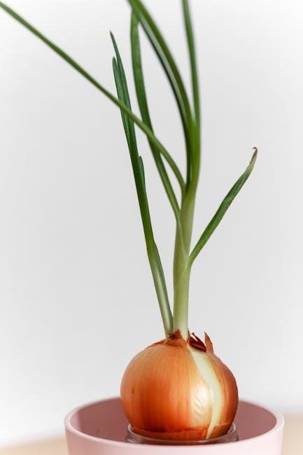
Onion root tips are excellent for studying mitosis due to their rapid cell division in the meristematic tissue. This tissue produces a high number of dividing cells, allowing easy observation of all cell cycle stages in a single preparation. The cells are large, organized in concentric layers, and simple to stain, making chromosomes visible under a microscope. This practicality makes onion root tips a widely used model in educational labs for demonstrating mitosis effectively.
1.3. Objectives of the Lab Activity
The primary objectives of this lab are to observe and identify the stages of mitosis in onion root tip cells. Students aim to understand the cell cycle, including interphase, prophase, metaphase, anaphase, telophase, and cytokinesis. Additional goals include mastering slide preparation, staining, and microscopy techniques. By analyzing the distribution of cells in each phase, students can infer the duration spent in each stage. This activity fosters practical skills in cellular biology and enhances understanding of mitosis through hands-on experimentation and data analysis.
Materials and Equipment Needed
Key materials include onion root tips, microscopes, glass slides, cover slips, stain solutions, and dissecting tools. Essential equipment comprises microscopes, slides, and basic laboratory tools;
2.1. List of Required Materials
- Microscope: Essential for observing cells at high magnification.
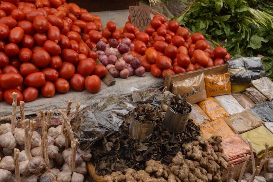
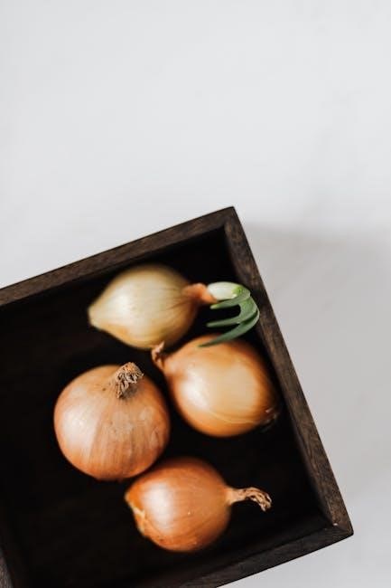
- Onion root tips: Provide actively dividing cells for mitosis observation.
- Glass slides and cover slips: For preparing and mounting cell samples.
- Sharp razor or scalpel: Used to cut thin root tip sections.
- Staining solutions: Such as methylene blue or toluidine blue for cell visibility.
- Carnoy’s fixative: Preserves cells and stops mitosis in its tracks.
- Forceps and dropper: For handling root tips and adding reagents.
- Water and blotting paper: For rinsing and drying slides.
Having all these materials ensures a smooth and successful mitosis observation experiment.
2.2. Preparation of the Microscope and Accessories
Before starting the lab, ensure the microscope is properly set up. Clean the lens with lens paper and adjust the stage clips. Plug in the light source and adjust the brightness. Place a drop of immersion oil on the stage for higher magnification. Focus the objective lens and condenser to achieve clarity. Prepare slides, cover slips, and other accessories nearby. Ensure all tools are within reach to streamline the process. Proper preparation of the microscope and accessories is crucial for clear cell observation and accurate results.
Step-by-Step Procedure for the Lab
Prepare the onion root tip slide, stain the cells, and observe under the microscope. Identify and record the stages of mitosis, ensuring accurate data collection for analysis.
3.1. Preparing the Onion Root Tip Slide
Begin by cutting a small section of the onion root tip and placing it on a microscope slide. Add a few drops of water to the root tip to help soften the tissue. Using a scalpel or sharp object, gently mash the root tip to release the cells. Apply a drop of toluidine blue or methylene blue stain to the slide to enhance chromosome visibility. Use a cover slip to flatten the cells further, ensuring even distribution. Allow the slide to air-dry before proceeding to observation under the microscope.
3.2. Staining the Cells for Better Visibility
After preparing the slide, a drop of toluidine blue or methylene blue stain is applied to the onion root tip cells. These dyes bind to the DNA, making the chromosomes more visible under the microscope. Gently heat the slide using a low-temperature flame or let it sit for a few minutes to allow the stain to set. Avoid over-staining, as it can obscure cell details. If necessary, rinse the slide with distilled water to remove excess dye, ensuring optimal visibility of the cell structures during observation.
3.3. Observing the Cells Under the Microscope
Place the prepared slide under the microscope and focus on the cells using low magnification (4x or 10x objective). Locate the root tip, where meristematic cells are densely packed. Switch to high magnification (40x) for detailed observation. Scan the slide systematically to identify cells in various stages of mitosis; Note the distinct features of each phase, such as chromatin condensation or chromosome alignment. Adjust lighting and focus as needed to enhance visibility and accurately record observations for further analysis and identification of mitotic stages.
3.4. Identifying Stages of Mitosis
Under the microscope, identify cells in different mitotic stages based on visible features. Cells in interphase show no dividing characteristics, while prophase cells display condensed chromosomes and a dissolving nucleus. In metaphase, chromosomes align at the cell center. During anaphase, sister chromatids separate to opposite poles. Telophase cells show re-forming nuclei, and cytokinesis reveals cell plate formation in plants. Record observations systematically to analyze the distribution of cells across stages for accurate data collection and interpretation.

Phases of Mitosis Observed in Onion Root Tips
The onion root tip cells exhibit distinct mitotic phases, including interphase, prophase, metaphase, anaphase, telophase, and cytokinesis, each with visible chromosomal and cellular changes under a microscope.
4.1. Interphase
Interphase is the longest stage of mitosis, where the cell grows, replicates its DNA, and prepares for cell division. During this phase, the nucleus is intact, and chromosomes are unwound, appearing as thin, thread-like structures. The cell performs normal metabolic functions, synthesizes proteins, and replicates organelles. Since interphase occupies about 90% of the cell cycle, most cells observed in the onion root tip will be in this phase, making it the most common stage seen under the microscope.
4.2. Prophase
Prophase marks the beginning of mitosis, where the chromatin condenses into visible chromosomes. The nucleolus disappears, and the nuclear envelope breaks down. Microtubules form the mitotic spindle, attaching to centrioles at the cell poles. This phase prepares the cell for chromosome alignment, ensuring proper distribution to daughter cells. Prophase is shorter than interphase but crucial for the accuracy of mitosis. In onion root tips, prophase cells show distinct chromosomal condensation and spindle formation, making this stage identifiable under the microscope.
4.3. Metaphase
Metaphase is the second stage of mitosis, where chromosomes align at the metaphase plate, an imaginary plane equidistant from the two poles of the cell. Spindle fibers attach to sister chromatids, ensuring proper alignment. This stage ensures each daughter cell will receive an identical set of chromosomes. In onion root tips, metaphase cells are easily identifiable due to their well-defined chromosome arrangement. This brief stage is critical for ensuring genetic continuity and accuracy in cell division.
4.4. Anaphase
Anaphase is a critical phase where sister chromatids separate and are pulled to opposite poles of the cell by spindle fibers. This ensures each daughter cell receives identical genetic material. In onion root tips, this stage is brief but vital for genetic continuity. The separation is essential for accurate distribution of chromosomes, making Anaphase a key step in mitosis.
4.5. Telophase
Telophase marks the beginning of the transition to cytokinesis. Chromosomes, now at opposite poles, start to decondense and nuclear envelopes reform around each set. Nucleoli reappear, and the cytoplasm prepares for division. In onion root tips, this phase is crucial as it ensures genetic material is correctly partitioned. Observing telophase helps confirm the cell cycle’s progression, with plant cells beginning to form a new cell wall. This phase is essential for maintaining genetic stability and continuity in dividing cells.
4.6. Cytokinesis
Cytokinesis follows telophase, completing cell division. In plant cells, a cell plate forms at the center, gradually developing into a new cell wall. This process divides the cytoplasm and organelles equally between the two daughter cells. Observing cytokinesis in onion root tips highlights the physical separation of cells, ensuring each daughter cell receives identical genetic material. This final stage is crucial for plant growth, as it allows root tips to maintain rapid cell division and structural integrity, ensuring proper tissue development and function.
Data Collection and Analysis
Students count cells in each mitotic phase and calculate percentages to determine the duration spent in each stage. This analysis provides insights into the cell cycle dynamics.
5.1. Counting Cells in Each Phase
Students systematically count cells in interphase, prophase, metaphase, anaphase, and telophase. Using a microscope, they tally cells in each stage, ensuring accuracy by examining multiple fields of view. Proper counting techniques are emphasized to avoid miscounting. The counts are recorded for further analysis, providing a clear distribution of cells across the mitotic phases.
5.2. Calculating the Percentage of Cells in Each Phase
To determine the percentage of cells in each phase, divide the number of cells in a specific phase by the total number of cells counted. Multiply the result by 100 to obtain the percentage. This calculation helps estimate the relative time cells spend in each phase. For example, a higher percentage in interphase indicates most cells are preparing for division. Accurate calculations provide insights into the cell cycle dynamics, allowing students to visualize how mitosis progresses and understand its biological significance.
5.3. Determining the Time Spent in Each Phase
Calculating the time spent in each phase involves using the percentages obtained. Since mitosis is a continuous process, the percentage of cells in each phase reflects the duration spent there. For instance, a higher percentage in interphase suggests it lasts longer. By comparing percentages across phases, students can infer the relative time each stage occupies in the cell cycle. This analysis enhances understanding of mitosis timing and its role in cellular processes.
Challenges and Common Mistakes
Common challenges include difficulty identifying mitotic stages, improper staining techniques, and miscounting cells. These issues can lead to inaccurate data and misinterpretation of results.
6.1. Difficulty in Identifying Stages
Identifying mitotic stages in onion root tips can be challenging due to the similarity between interphase and early prophase cells. Overlapping or crowded cells may obscure visibility, making it hard to distinguish stages accurately. Additionally, poor staining can reduce chromosomal detail, complicating phase identification. Students often confuse interphase cells with those in prophase due to chromatin condensation. Proper training and comparison with diagrams are essential to improve accuracy and avoid misclassification of cell stages during observations.
6.2. Incorrect Staining Techniques
Incorrect staining is a common issue in the onion root tip mitosis lab, leading to poor visibility of chromosomes. Overstaining can obscure cellular details, while understaining makes chromatin barely visible. Using expired or diluted stains also reduces effectiveness. Proper staining involves precise timing and concentration to ensure cells are adequately dyed without damaging the sample. Following the protocol carefully is crucial to achieve clear, distinguishable chromosomal structures under the microscope, enabling accurate phase identification during mitosis observations in the lab report.
6.3. Miscounting Cells
Miscounting cells is a common challenge in the onion root tip mitosis lab, often due to human error or overlapping cells in the field of view. Students may inadvertently count the same cell multiple times or miss cells entirely, leading to inaccurate data. To mitigate this, a systematic counting method should be employed, such as using a grid or tallying cells in a specific pattern. Double-checking counts or using digital tools can also improve accuracy and ensure reliable results for phase percentage calculations.

Interpretation of Results
Interpreting results involves analyzing cell cycle phases, comparing observed data with theoretical expectations, and understanding the duration cells spend in each stage of mitosis.
7.1. Understanding the Cell Cycle
The cell cycle consists of interphase and mitosis. Interphase is the longest stage where cells grow, replicate DNA, and prepare for division. Mitosis is divided into prophase, metaphase, anaphase, and telophase, each with distinct chromosomal changes. Cytokinesis follows, splitting the cytoplasm and forming two daughter cells. By observing onion root tip cells, students can visually track these stages, gaining insights into how cells regulate growth and division, essential for understanding tissue repair, development, and cancer biology.
7.2. Relating Observations to Theoretical Concepts
Observing onion root tip cells aligns with theoretical concepts of the cell cycle, where most cells are in interphase, and few in mitosis. This reflects the brief duration of mitosis. Calculating cell percentages in each phase supports theories about cell cycle regulation and time allocation. These practical findings validate textbook explanations, demonstrating how experimental data connects to broader biological principles. This exercise bridges the gap between theory and observation, reinforcing understanding of mitotic processes and their significance in growth and development.
Drawing Conclusions
The onion root tip mitosis lab effectively demonstrates cell division stages, validating theoretical concepts. Observations reveal the majority of cells in interphase, highlighting mitosis’s brief duration and its role in growth.
8.1. Summary of Key Findings
The onion root tip mitosis lab revealed that most cells were in interphase, with fewer in prophase, metaphase, anaphase, or telophase. This indicates that mitosis is a brief process. Observations confirmed that rapid cell division in root tips provides an ideal model for studying mitosis. Data collection showed varying percentages of cells in each phase, allowing estimation of the time spent in each stage. The lab successfully demonstrated the cell cycle’s progression and reinforced theoretical concepts through practical observation and analysis.
8.2. Implications for Cell Biology
The study of mitosis in onion root tips provides insights into fundamental cell division mechanisms. Observing these cells helps understand how genetic material is duplicated and distributed, ensuring cellular continuity. This lab highlights the universality of mitosis across eukaryotic organisms, offering a simple model to explore complex biological processes. The findings also underscore the importance of interphase, where most cellular activities occur, and how brief mitosis is relative to the cell cycle. Such studies are crucial for advancing research in genetics, cancer biology, and plant development.
Extensions and Variations of the Lab
Explore mitosis in other tissues, such as animal cells, or compare plant and animal cell division. Additional experiments could involve testing environmental factors affecting mitosis rates.
9.1. Using Different Plant Tissues
Exploring mitosis in various plant tissues, such as shoot apices or garlic cloves, provides a broader understanding of cell division. These tissues, like onion root tips, contain meristematic cells with high division rates. Students can compare mitotic activity across tissues, noting differences in cell arrangement and division stages. This extension enhances observational skills and reinforces concepts of plant growth patterns. Additionally, experimenting with alternative tissues can address challenges in obtaining onion roots or offer varied perspectives on mitotic processes in plants.
9.2. Comparing Plant and Animal Cells
Comparing plant and animal cells during mitosis highlights key differences in cell division. Plant cells form a cell plate during cytokinesis, while animal cells undergo centripetal contraction. Observing both types allows students to note variations in cell wall structure and division mechanisms. This extension enriches understanding of cellular diversity and reinforces mitotic concepts. It also challenges students to adapt lab techniques for animal tissues, such as human skin cells or frog tadpole tail cells, broadening their practical skills in cell biology.

Frequently Asked Questions
This section addresses common queries about the onion root tip mitosis lab, such as why onion roots are used, improving slide preparation, and troubleshooting issues.
10.1. Why Are Onion Root Tips Used?
Onion root tips are used because they contain rapidly dividing meristematic cells, making mitosis stages easily observable. Their cell walls and large chromosomes aid visibility under a microscope. The root tips are simple to prepare for slides, and their active growth ensures various stages of mitosis are present. This makes them an ideal model for studying cell division in plant cells, providing a practical and effective learning tool for students.
10.2. How to Improve Slide Preparation?
To improve slide preparation, ensure the onion root tip is fresh and actively growing. Carefully slice thin sections to maintain cell integrity. Use a clean slide and cover slip to prevent contamination. Gently squash the tissue to spread cells evenly without tearing. Apply the correct staining technique to enhance chromosome visibility. Avoid over-staining, as it can obscure details. Properly fix and rinse the sample to maintain cell structure. Handling the specimens gently prevents distortion, ensuring clear observation under the microscope.
10.3. What If No Dividing Cells Are Observed?
If no dividing cells are observed, it may indicate issues like improper staining, incorrect slicing, or insufficient cell activity. Ensure the root tip is fresh and actively growing. Check staining procedures for accuracy and visibility. Re-examine the slide under the microscope, focusing on areas with higher cell density. If the problem persists, consider preparing a new slide or selecting a different root tip. Proper technique and sample quality are crucial for observing mitotic cells effectively.

Answer Key for the Lab Report
This section provides correct identifications of mitosis stages, sample calculations for cell percentages, and expected results to help students accurately analyze and interpret their lab findings.
11.1. Correct Identification of Mitosis Stages
The answer key provides detailed descriptions and illustrations of each mitosis stage, from interphase to cytokinesis. It helps students accurately identify and label cells, ensuring precise recognition of chromosomal behavior and cellular changes. This guide is essential for avoiding common misconceptions and improving observational skills under the microscope.
11.2. Sample Calculations for Cell Percentages
Students count cells in each phase of mitosis, calculate percentages, and compare findings. For example, if 50 cells are observed and 10 are in metaphase, the percentage is (10/50) × 100 = 20%. This helps estimate the time cells spend in each phase. The mitotic index, calculated as (number of dividing cells/total cells) × 100, provides insights into cellular activity. These calculations enhance understanding of the cell cycle dynamics and validate observations.
11.3. Expected Results and Analysis
Expected results show most cells in interphase, with fewer in prophase, metaphase, anaphase, and telophase. Calculations reveal interphase occupies ~80-90% of cells, while mitotic phases account for 10-20%. Analysis confirms that interphase lasts longer than other stages, aligning with the cell cycle’s duration. The mitotic index, calculated as (dividing cells/total cells) × 100, typically ranges from 5-15%. These findings support theoretical concepts of cell cycle dynamics and provide practical insights into mitosis in plant cells.
Final Thoughts and Reflections
This lab provided hands-on experience in observing mitosis, enhancing understanding of cell division and practical skills in slide preparation and microscopy, offering insights into cellular processes.
12.1. Learning Outcomes
Students gained hands-on experience in observing and identifying mitosis stages in onion root tips. They learned to prepare microscope slides, stain cells, and calculate the percentage of cells in each phase. This lab enhanced understanding of the cell cycle, emphasizing interphase’s dominance. Practical skills in microscopy and data analysis were developed, fostering critical thinking and scientific inquiry. These outcomes provide a foundation for advanced studies in cell biology and related fields, highlighting the importance of mitosis in cellular processes.
12.2. Practical Skills Gained
Students developed essential laboratory skills, including slide preparation, cell staining, and microscopy techniques. They learned to identify mitotic stages accurately and interpret cellular structures. Practical experience in data collection and analysis enhanced their ability to calculate cell percentages and draw conclusions. Additionally, the lab improved their critical thinking and scientific inquiry skills, enabling them to connect observations with theoretical concepts. These practical skills are invaluable for future scientific investigations and experimentation in biology.
12.3. Suggestions for Future Experiments
Future experiments could explore the effects of environmental factors, such as temperature or chemicals, on mitosis in onion root tips. Comparing mitosis in different plant tissues or between plant and animal cells could provide deeper insights. Additionally, investigating the role of specific genes or proteins in cell division could enhance understanding. Long-term observations of cell cycle duration and variability could also yield valuable data. These extensions would further enrich students’ understanding of cellular biology and experimental design.